

Eye Bag Surgery: Why Removing Fat Is Usually the Wrong Answer
By Dr. Rushapol Sdawat, MD — Thai Board-certified Plastic and Reconstructive Surgeon, Medical Licence No. 17689
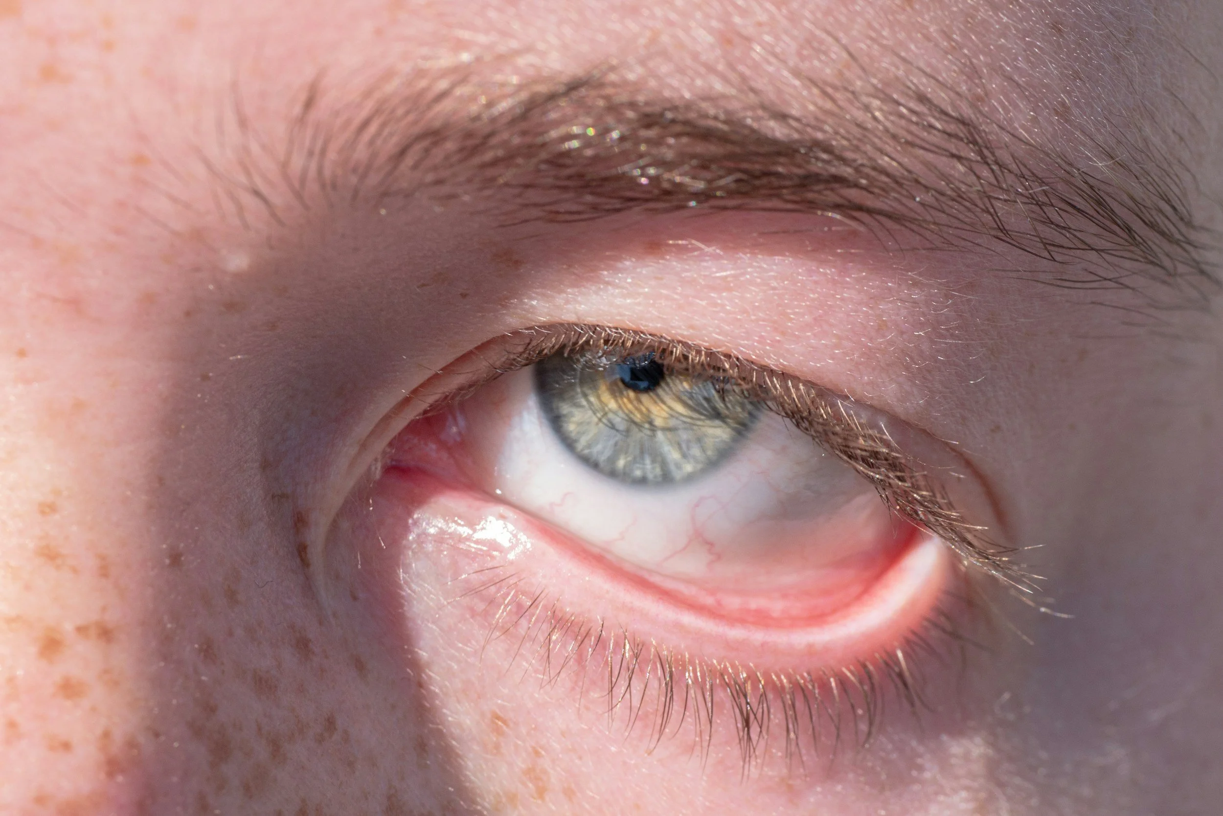
You have taken the same photograph of yourself twice — once in flat indoor light, where you look fine, and once outside at midday, where two grey crescents appear under your eyes that you do not recognise. You have concluded that you have bags and that a surgeon should take them out.
That conclusion is understandable and it is usually half wrong. What you are looking at in bright overhead light is very often not a lump of fat but a shadow — cast by a step between the lower eyelid and the cheek. Sometimes the step exists because fat has come forward. Just as often it exists because the ground beneath has fallen away, and the fat sitting in front of it is the only thing stopping the area from looking skeletal.
This distinction is the whole operation. Get it wrong in a thirty-eight-year-old and you will produce a lower lid that looks tidy at six weeks, hollow at eight months and noticeably older at five years — and there is no good way back.
What you are actually looking at is a shadow, not a bag
Do this now. Look at yourself with a light source above you, then hold a phone torch at chin height and look again. If the crescents largely vanish when lit from below, they were shadows created by a contour step, not by volume sitting on the surface.
Then lie flat and look in a hand mirror. Fat herniating through a lax orbital septum moves with gravity and will visibly soften when you are supine. A hollow does not move; it stays exactly where it was.
Most patients have some of both. The proportion is what determines whether anything should be removed at all.
Why the eye bag is a lid–cheek junction problem
The lower eyelid does not end at the lash line and start again at the cheek. Between them runs the lid–cheek junction, and its shape is set by three things: the position of the orbital rim bone underneath, the orbicularis retaining ligament that tethers soft tissue to that rim, and the descent of the midface fat over time.
The tear trough is the medial part of that groove — the depression running down and outwards from beside the nose. It deepens with age because the retaining ligament holds the skin down at a fixed line while the cheek fat below slides away from it, and because orbital fat above the line pushes forward. You end up with a bulge above a groove, and the eye reads as tired.
Nothing about that description says "remove tissue." Two of the three mechanisms are loss and descent. Only one is excess.
The case against taking fat out
Here is what happens over the following decade if you remove orbital fat generously from someone in their thirties or forties.
At six weeks the lid looks smooth and the patient is delighted. Over the next six to twelve months the residual oedema resolves and the true volume becomes visible. The orbital rim starts to show as a hard line. Light no longer reflects off a convex lower lid; it falls into a concave one. The eye appears to sink, the sclera below the iris can look more exposed, and the face reads as gaunt rather than rested — an appearance sometimes described as a skeletonised or hollowed lower lid.
And it is very hard to reverse. Correcting a post-surgical hollow means fat grafting into a scarred, thin, mobile, unforgiving tissue plane where lumps and irregularities show through skin under a millimetre thick, or placing implants over the rim. Both are real operations with real failure rates. Neither reliably restores what was there. This is one of the few things in facial surgery where the revision is genuinely worse than the primary operation, and it is why I will argue with a patient who arrives asking me to "take all of it out."
Fat removal is not always wrong. In an older patient with genuinely large, long-standing herniated pads and a well-supported rim, conservative reduction is appropriate and works. The word doing the work is conservative.
Arcus marginalis release and fat repositioning
The alternative is to stop treating the fat as waste and start treating it as filler that is already in the right postcode.
The arcus marginalis is the dense fibrous line where the orbital septum fuses to the periosteum at the orbital rim. It is the reason a bulge stops abruptly at a groove instead of blending into the cheek. Releasing it — dividing that fusion along the rim — removes the tether.
Once released, the herniated orbital fat can be mobilised as pedicled flaps, brought down over the rim into the space where the tear trough was, and secured there. The bulge above the groove and the depression below it are addressed by the same manoeuvre, using the patient's own tissue, with the fat retaining its blood supply rather than being grafted blind.
I usually approach this from behind the lid, through the conjunctiva, which leaves no external scar and — crucially — does not divide the orbicularis oculi muscle or create a skin scar that can contract downwards.
Why the lift often comes from above, not from cutting the lid
There is a habit in this field of treating the lower eyelid as the site of the problem because it is the site of the complaint. Frequently it is not.
If the midface has descended, the lid–cheek junction lengthens and a step appears at the rim. You can chase that step from the lid all day. What actually shortens it is re-supporting the cheek — elevating and fixing the midface soft tissue so that it meets the lid again from below. That may be a midface suspension performed through the same lower lid approach, a temporal or extended deep plane dissection, or structural fat grafting to the upper cheek rather than to the lid itself.
Adding volume above the groove is often safer than working within it. The skin is thicker, the plane is more forgiving, and there is no lower lid margin to pull on. The most reliable way to avoid pulling a lower lid down is to not put tension on it in the first place.
Snap-back, distraction and the tests that decide whether I operate
Before any of the above, I assess lid support, and this takes about twenty seconds.
Snap-back test. The lower lid is pulled down away from the globe and released without the patient blinking. A normal lid snaps back against the eye immediately. The longer it takes, the more laxity is present.
Distraction test. The lid is pulled forward away from the eye between thumb and finger. Distraction of more than 2 mm from the globe is abnormal — roughly, 2–4 mm mild, 4–6 mm moderate, over 6 mm severe.
Negative vector. Viewed from the side, if the most projecting point of the cheek sits behind the front of the cornea, the lid has no bony shelf to rest on. These patients are at markedly higher risk of lid retraction and need canthal support and a very conservative approach.
If you have laxity and I do not add support, the lid will very likely sit lower afterwards. Canthal support means either a canthopexy — tightening and re-suspending the lateral canthal tendon without dividing it — or a canthoplasty, where the tendon is divided, shortened and re-fixed to the inner orbital rim. Canthoplasty is more powerful and less forgiving.
Support is not free. In the 892-case blepharoplasty series published in JAMA Facial Plastic Surgery in 2013, intraoperative canthopexy was associated with chemosis — conjunctival swelling — in 45% of cases compared with 25.5% without it. Chemosis is temporary and treatable, but it prolongs recovery, and I would rather you heard that number from me than discovered it at day ten.
Transconjunctival versus transcutaneous
TransconjunctivalTranscutaneous (subciliary)IncisionInside the lid, behind the lashesSkin, 1–2 mm below the lash lineExternal scarNoneFine line, usually settles wellOrbicularis muscleNot dividedDivided or elevatedCan excess skin be removed?No — needs a separate skin pinch or laserYesAccess for fat repositioningGoodGoodRisk of lid retraction/ectropionLowerHigher — the classic causeSuitsMost patients under about 55; any lid with laxityGenuine significant skin excess with a well-supported lidSuturesUsually none or dissolvingRemoved day 5–7
The reason transconjunctival access has become my default is not fashion. It is that the middle lamella — septum and muscle — is the layer that scars and contracts, and going behind the lid leaves it largely alone.
Ectropion is the complication that defines this operation
Ectropion is outward turning of the lower lid away from the eye. Its milder relatives are lid retraction, in which the margin sits lower than it should and white sclera shows below the iris, and rounding of the lateral canthus, in which the eye loses its almond shape and looks subtly wrong in a way patients cannot name.
They happen for three reasons: unrecognised pre-existing laxity, over-resection of lower lid skin, and scar contracture in the middle lamella after a transcutaneous approach. All three are decisions made before or during the operation, not accidents afterwards.
Rates in careful hands are low — one large series reported symptomatic lid malposition in 0.4% of 2,007 transcutaneous lower blepharoplasties over 30 years. That figure describes a meticulous single-surgeon practice with proper patient selection. It does not describe what happens when a lid with 5 mm of distraction and a negative vector has 4 mm of skin excised in a high-volume list.
The rest of the risk list is real and belongs on the table: haematoma, infection, prolonged chemosis, dry eye and grittiness, temporary blurred vision, persistent oedema for two to three months, visible or palpable irregularity of repositioned fat, asymmetry between sides, numbness, and — rarely — orbital haemorrhage behind the eye, reported at roughly one in 2,000 cosmetic periocular operations with permanent vision loss at roughly one in 22,000. And, as with all facial surgery, a result you are technically fine with but emotionally disappointed by.
Who I decline
I do not operate on a lower lid where the dominant finding is hollowing and the patient still wants fat removed after I have explained why. I decline where lid laxity is significant and the patient refuses canthal support. I decline in active thyroid eye disease that has not been stable for at least six months, and in significant untreated dry eye. I am cautious in anyone who has had previous lower lid surgery, laser resurfacing or a mid-face lift, because the middle lamella is already scarred.
And I will say the unhelpful thing plainly: for a good many people in their thirties with faint shadows, the correct answer is no surgery at all. Improve sleep and salt, treat allergic rhinitis if it is present, consider a small volume of well-placed filler or fat above the groove, and reassess in two years. Surgery on a lower lid is not a starting point. It is a decision you make once.
When to seek care
Emergency — hospital immediately, day or night. Sudden severe deep pain behind the eye, especially with rapidly increasing swelling and a tight, forward-bulging eye. Any loss of vision, a dark curtain, or new double vision. This pattern suggests orbital haemorrhage and there is a short window in which pressure can be released. Do not wait until morning, do not board a flight, and do not message a coordinator and go back to bed.
Same-day review. Fever above 38 degrees. Spreading redness, heat and increasing pain after day three, when things should be improving. Pus or a discharging wound. Bleeding that will not stop with gentle pressure. Sudden marked asymmetry that was not there yesterday. A lid that is visibly pulling away from the eye.
Within a few days. A gritty, burning or watering eye not settling with lubricant drops. Persistent conjunctival swelling that looks like a blister of clear jelly on the white of the eye. A lower lid margin sitting lower than it did last week. A palpable lump where fat was repositioned that is not softening by week six.
If you are still in Bangkok, contact the hospital directly rather than whoever arranged the trip. If you are already home in Australia or New Zealand, present locally and bring your operation note. Do not delay because your surgeon is overseas — and be aware that Australian private health insurance generally does not cover procedures performed overseas and may not cover the follow-up either, and that in New Zealand, NZAPS states ACC does not cover injuries from surgery performed overseas.
What I would want you to ask any surgeon offering this
Ask them to tell you, looking at your face, how much of what you see is protruding fat and how much is hollow. Ask what they intend to do with the fat — remove it, or move it. Ask whether they have tested your lid laxity and what the result was. Ask what they will do about lid support and why. And ask what their plan is if, at eight months, the lid looks hollow.
If the answer to the first question is a number they have not measured and the answer to the last is silence, keep looking.
I am a Thai Board-certified plastic and reconstructive surgeon practising at Intrarat Hospital, Bangkok — Medical Licence No. 17689, verifiable on the Medical Council of Thailand register at checkmd.tmc.or.th. Diploma of the Medical Council of Thailand certifying proficiency in Plastic Surgery (13 July 2000); full member of The Society of Plastic and Reconstructive Surgeons of Thailand since 2001; ISAPS member since 2008. International training includes the AO Foundation course in advances in rhinoplasty and facial osteotomy, the International Plastic Surgery Training Center (malarplasty and angle reduction) and the safe use of laser-assisted lipolysis at Goldman Butterwick & Associates, San Diego. I hold Thai specialist certification and am not registered with Ahpra or the Medical Council of New Zealand.
This article is provided for general information and education. It is not medical advice and cannot substitute for individual assessment by a qualified practitioner who knows your history. Surgery carries risk, including risk of serious complications and of outcomes that do not meet expectations. Results vary between patients.
